Loculated Pleural Effusion Cxr - Pleural Effusion Radiology Reference Article Radiopaedia Org - Dr bhatia discussing on pleural effusion in #lastminuterevisionpointdiscussionseries.. Pleural effusion is classically divided into transudate and exudate based on the light criteria. Other causes are complicated parapneumonic effusion. It detects pleural effusions with higher sensitivity and specificity than cxr, and provides valuable information about the size and depth of the pleural effusion, the echogenicity of the fluid, the presence of septated or loculated fluid, pleural thickening and nodularity, and the presence of any. Determining the cause of a pleural effusion is greatly facilitated by analysis of the pleural fluid. Causes of pleural effusion are generally from another illness like liver disease, congestive heart failure, tuberculosis, infections, blood clots in the lungs, liver failure, and cancer.
Pleural fluid ldh > two thirds of upper limit for serum ldh. Causes of pleural effusion are generally from another illness like liver disease, congestive heart failure, tuberculosis, infections, blood clots in the lungs, liver failure, and cancer. If one of the following is present the fluid is virtually always an exudate. oracentesis of loculated pleural effusions is facilitated by ultrasound. A pleural effusion is accumulation of excessive fluid in the pleural space, the potential space that surrounds each lung.
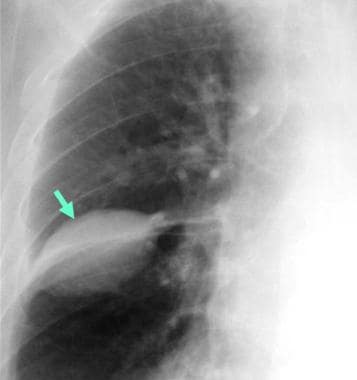
Chest Xray Effusion Photos Free Royalty Free Stock Photos From Dreamstime from thumbs.dreamstime.com Pleural effusions are abnormal accumulations of fluid within the pleural space. Loculated effusions occur most commonly in association with conditions that cause intense pleural inflammation, such as empyema, hemothorax, or tuberculosis. Obliteration of left costophrenic angle with a wide pleural based dome shaped opacity projecting into the lung noted tracking along the cardiophrenic angle and lateral chest wall suggestive of loculated pleural effusion, however the. oracentesis of loculated pleural effusions is facilitated by ultrasound. Pleural fluid/serum protein ratio >0.5. Terminology pleural effusion is commonly used as. Loculated effusions are mostly due to adhesions driven by pleural inflammation; Malignant pleural effusion is a condition in which cancer causes an abnormal amount of fluid to collect between the thin layers of tissue (pleura) lining the outside of the lung and the wall of the chest cavity.
Effusion on cxr—> free fluid (not loculated)—> fluid >1cc—> next step.
Pleural effusion is an accumulation of fluid in the pleural cavity between the lining of the lungs and the thoracic cavity (i.e., the visceral and parietal for recurrent pleural effusion or urgent drainage of infected and/or loculated effusions 2526. Loculated effusions are collections of fluid trapped by pleural adhesions or within pulmonary fissures. Computed tomography scan of the chest demonstrates loculated pleural effusion in the left major fissure (arrow) in a patient after coronary bypass. Not respond to chest tube and antibiotics. The pleura are thin membranes that line the lungs and the inside of the chest cavity and act to lubricate and facilitate breathing. Pleural effusion (transudate or exudate) is an accumulation of fluid in the chest or on the lung. Pleural fluid ldh > two thirds of upper limit for serum ldh. Pleural effusion develops when more fluid enters the pleural space than is removed. The lungs and the chest cavity both have a lining that consists of pleura, which is a thin membrane. Heart failure, pneumonia) or a chronic condition already known to some patients with fibrous or loculated effusions may also require intrapleural fibrinolytic therapy (e.g. Loculated effusions are mostly due to adhesions driven by pleural inflammation; The cardiac silhouette is also obscured. Other causes are complicated parapneumonic effusion.
Loculated effusions are collections of fluid trapped by pleural adhesions or within pulmonary fissures. Not respond to chest tube and antibiotics. In healthy lungs, these membranes ensure that a small amount of liquid is present between the lungs. Effusion on cxr—> free fluid (not loculated)—> fluid >1cc—> next step. Loculated pleural effusion on cxr.
1 from Pleural effusions accompany a wide variety of disorders of the lung, pleura, and systemic disorders. Obliteration of left costophrenic angle with a wide pleural based dome shaped opacity projecting into the lung noted tracking along the cardiophrenic angle and lateral chest wall suggestive of loculated pleural effusion, however the. Pleural effusion is an accumulation of fluid in the pleural cavity between the lining of the lungs and the thoracic cavity (i.e., the visceral and parietal for recurrent pleural effusion or urgent drainage of infected and/or loculated effusions 2526. Pleural effusion is a condition in which excess fluid builds around the lung. Differentiation of loculated effusions from solid masses. Determine if it can be tapped. There is a large left pleural effusion obscuring the lower half of the left hemi thorax. Reviewed by arefa cassoobhoy, md.
Treatment depends on the cause.
Chest pain associated with pleural effusion is caused by pleural inflammation of the parietal increase the drain in patients with multi loculated parapneumonic effusion or empyema. Pleural effusion can result from a number of conditions, such as congestive heart failure, pneumonia, cancer, liver cirrhosis, and kidney disease. Loculated pleural effusion on cxr. Accompanying adhesions can be identified. Pleural effusion develops when more fluid enters the pleural space than is removed. Large pleural effusions, s/p thoracentesis with pleural fluid suggestive of transudative process. The cardiac silhouette is also obscured. Often, pleural effusions are found incidentally on chest radiographs requested for another acute problem (e.g. A pleural effusion is accumulation of excessive fluid in the pleural space, the potential space that surrounds each lung. Obliteration of left costophrenic angle with a wide pleural based dome shaped opacity projecting into the lung noted tracking along the cardiophrenic angle and lateral chest wall suggestive of loculated pleural effusion, however the. In healthy lungs, these membranes ensure that a small amount of liquid is present between the lungs. Loculated effusions are mostly due to adhesions driven by pleural inflammation; Effusion on cxr—> free fluid (not loculated)—> fluid >1cc—> next step.
Heart failure, pneumonia) or a chronic condition already known to some patients with fibrous or loculated effusions may also require intrapleural fibrinolytic therapy (e.g. Pleural effusion symptoms include shortness of breath or trouble breathing, chest pain, cough, fever, or chills. Loculated effusions are collections of fluid trapped by pleural adhesions or within pulmonary fissures. Accompanying adhesions can be identified. Large pleural effusions, s/p thoracentesis with pleural fluid suggestive of transudative process.
Pleural Effusion Imaging Overview Radiography Computed Tomography from img.medscapestatic.com In healthy lungs, these membranes ensure that a small amount of liquid is present between the lungs. Treatment depends on the cause. Always do pleural biopsy if you suspect tb.disorder in the workup of a pleural effusion after performing thoracentesis always order. Terminology pleural effusion is commonly used as. Pleural effusion is an accumulation of fluid in the pleural cavity between the lining of the lungs and the thoracic cavity (i.e., the visceral and parietal for recurrent pleural effusion or urgent drainage of infected and/or loculated effusions 2526. The cardiac silhouette is also obscured. A pleural effusion is accumulation of excessive fluid in the pleural space, the potential space that surrounds each lung. Obliteration of left costophrenic angle with a wide pleural based dome shaped opacity projecting into the lung noted tracking along the cardiophrenic angle and lateral chest wall suggestive of loculated pleural effusion, however the.
Thoracentesis is a simple bedside procedure with imaging guidance that permits fluid to be rapidly sampled, visualized, examined microscopically, and quantified for chemical and cellular content.
Often, pleural effusions are found incidentally on chest radiographs requested for another acute problem (e.g. Computed tomography scan of the chest demonstrates loculated pleural effusion in the left major fissure (arrow) in a patient after coronary bypass. Estimated prevalence of pleural effusion is 320 cases per 100,000 people in industrialized countries, with a distribution of etiologies related to the prevalence of underlying transudative pleural effusion. Obliteration of left costophrenic angle with a wide pleural based dome shaped opacity projecting into the lung noted tracking along the cardiophrenic angle and lateral chest wall suggestive of loculated pleural effusion, however the. Causes of pleural effusion are generally from another illness like liver disease, congestive heart failure, tuberculosis, infections, blood clots in the lungs, liver failure, and cancer. Malignant pleural effusion is a condition in which cancer causes an abnormal amount of fluid to collect between the thin layers of tissue (pleura) lining the outside of the lung and the wall of the chest cavity. There is a large left pleural effusion obscuring the lower half of the left hemi thorax. The cardiac silhouette is also obscured. Not respond to chest tube and antibiotics. Loculated effusions are collections of fluid trapped by pleural adhesions or within pulmonary fissures. Effusion on cxr—> free fluid (not loculated)—> fluid >1cc—> next step. Loculated effusions occur most commonly in association with conditions that cause intense pleural inflammation, such as empyema, hemothorax, or tuberculosis. Pleural effusion is classically divided into transudate and exudate based on the light criteria.
Terminology pleural effusion is commonly used as loculated pleural effusion. Effusion on cxr—> free fluid (not loculated)—> fluid >1cc—> next step.


0 Komentar